

Features
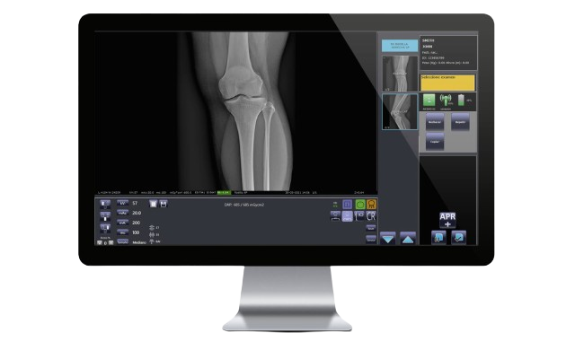
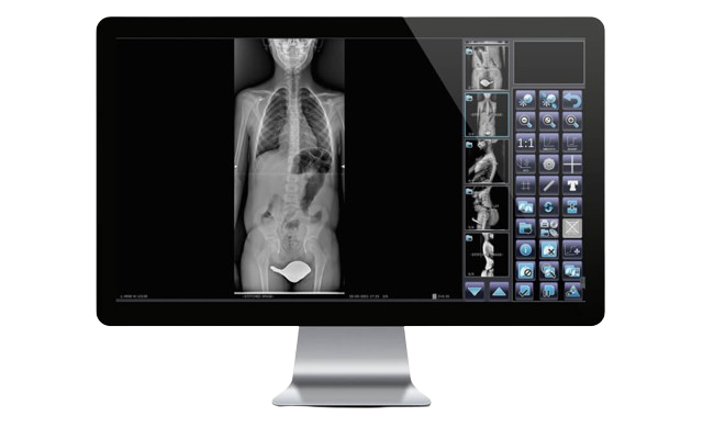
- Optimized image processing from the latest generation digital detector, thanks to Canon's CXDI NE and PRIMOS software incorporated in our fixed and mobile equipment, offering anatomical sharpness and image resolution never before achieved.
- Thanks to the preview possibility they can be accepted and sent immediately via cable and/or WiFi to the Hospital's image server (PACS) for analysis with advanced imaging tools on workstations or sent directly to consultations (DICOM) or exported to printers or external devices such as DVD or USB (in JPG and DICOM format).
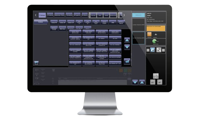
- Software that will make a difference in your service with images that hardly need to be adjusted thanks to their extremely high quality and consistency in each exposure. Save time and achieve a more efficient workflow while optimizing dose control for both the patient and the medical team. Achieve repeat-free diagnosis with the most advanced technology.
Acquisition
- Low Dose Fluoroscopy.
- High Quality Fluoroscopy (high quality).
- Digital Radiography (instant image).
- Fluoroscopy in Road Mapping mode (optional).
- Fluoroscopy in DSA mode: Remasking, Subtraction, Landmarking, vascular tracking and stenoisis (optional).
Optional
- Upgrade 20kW
- Laser locator.
- Integrated dosimeter.
- DSA vascular packete. In
- Automatic login.
- DICOM functions.
- Wi-Fi pedal.
- RFID card reader.
- Wireless trolley.
- Detector 30×30.
- Rotating anode.
Clinical Use
- Easily process and reconstruct images from the digital detector with state-of-the-art technology, thanks to the Canon CXDI NE and PRIMOS software included in our fixed and mobile equipment.
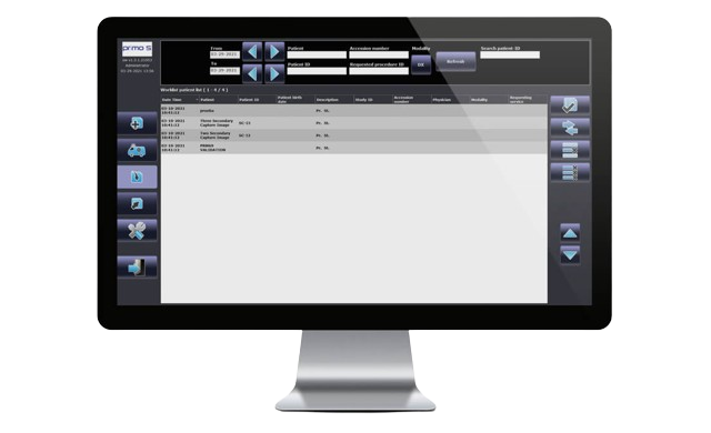
- Thanks to the possibility of immediate preview they can be accepted and sent via cable and/or WiFi to the Hospital's image server (PACS) for analysis and further processing with advanced imaging tools on workstations and finally be sent directly to consultations (DICOM) or exported to printers or external devices such as DVD or USB (in JPG and DICOM format).
- Software that will make a difference in your service with images that hardly need to be adjusted thanks to their extremely high quality and consistency in each exposure. Save time and achieve a more efficient workflow while optimizing dose control for both the patient and the medical team. Achieve repeat-free diagnosis with the most advanced technology.
Technical Specifications
Column / Table
- Motorized Table Tilting
- 90°. Speed at 6°/s
- Tube rotation
- ± 180°
- (SID Min-Max) Motorized DFP
- 110cm - 180cm
- Motorized column angulation
- ± 40°
- Collimator
- Automatic and Motorized with LED and laser light
- Collimator rotation (manual)
- ±45º (optional)
- Longitudinal movement detector Motorized
- Longitudinal movement detector Motorized
- Detector rotation
- Vertical/horizontal (40°/s)
- Overall dimensions and weight
- 226 x 235 x 189 cm, 1600Kg. (In horizontal position, minimum height, minimum SID, column 0º)
- Board Dimensions
- 225 x 81 cm
- Vertical movement of table top (variable height) Motorized
- From 38 to 148cm, speed 6cm/s
- Longitudinal movement Motorized board
- 120/60cm (other lower values on request), speed 8cm/s
- Motorized lateral movement
- ± 18cm, speed 6cm/s
- Equivalent filtration
- 0.55 mm Al at 100kVp
- Width of radiolucent area
- 60cm
- Material Board
- Carbon Fiber
- Maximum Patient Weight (IEC 60-601-1)
- 230Kg without limitation, 310Kg with restrictions
- Footrest width
- 40cm, supported weight 230Kg
Automatic Tomography
- Curvilinear tomography in one direction
- Three angles can be selected during installation from 8° to 40°.
- Thickness of the cutting layer
- 1.5mm (at 40°), 3mm (at 20°), 6mm (at 10°)
- Maximum Speed
- 0.5s for 10°, 1s for 20°, 2s for 40°.
- Fulcrum height
- From 0 to 300mm in steps of 1mm or 5mm. Automatic adjustment over the set height (increments and decrements) in the millimeters desired by the operator.
Motorized Retractable Compression Device (optional)
- Pallet compression
- Up to 200N (130N factory programmed) with a maximum column angulation of ±10°.
- Minimum distance to the board in compression
- 10cm
- Patient Safety System
- Inhibition of table top, spine and angulation movements during compression. In case of power failure on the table, if the patient is compressed, it can be released by a manual system.
Documentation
Related Products
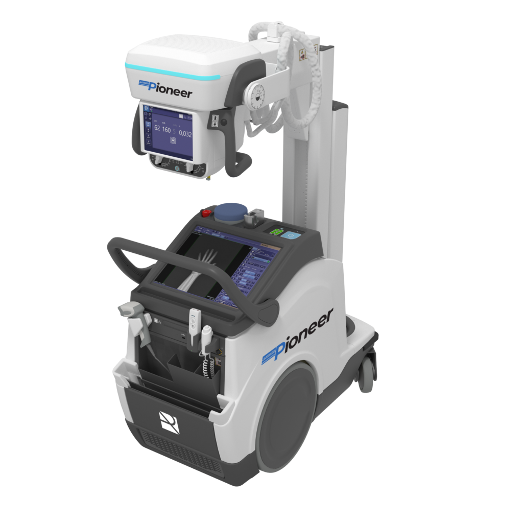
Digital mobile with energy recovery technology, in a lightweight, motorized and compact design that allows you to bring the diagnosis with the patient and more complicated environments such as ICU, neonatal centers, etc..
View product
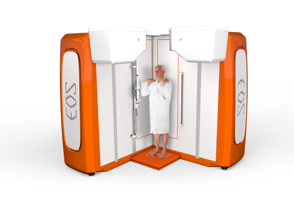
EOSedge proporciona, en un entorno abierto y confortable para el paciente, una evaluación global del sistema esquelético para facilitar la toma de decisiones terapéuticas cruciales.
Los modelos 3D y los parámetros clínicos 3D asociados ayudan a los médicos a evaluar las patologías musculoesqueléticas e identificar opciones de tratamiento para mejorar los resultados clínicos.
View product
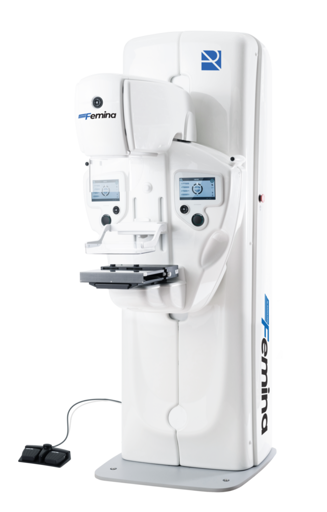
Analog mammography system designed for screening and diagnostic processes, with high frequency generator, Potter-Bucky system and a wide range of accessories for magnification, two-dimensional biopsies and precise positioning, guaranteeing high quality images and reliable diagnosis.
View product
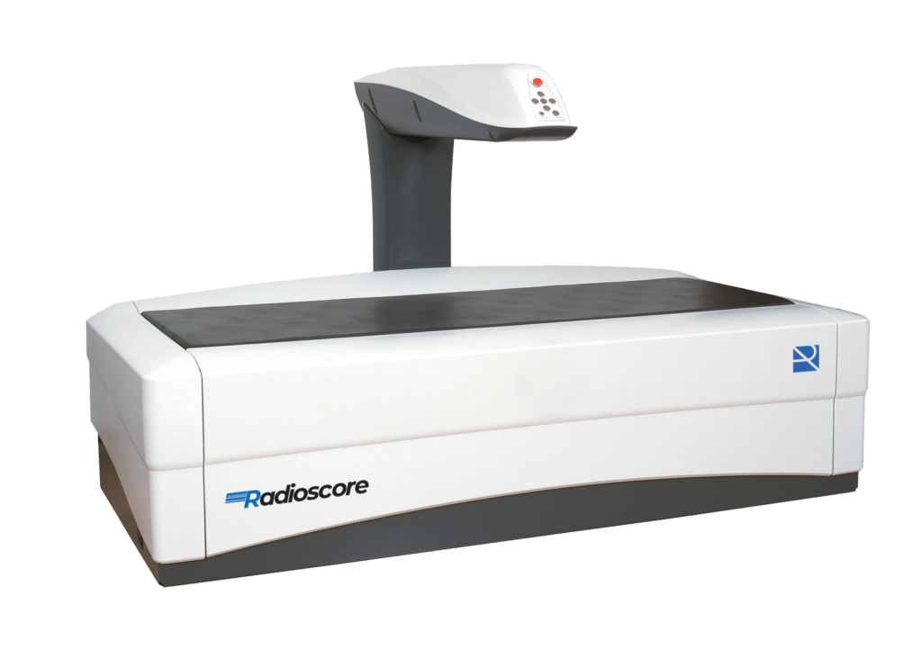
Pencil Beam analog technology DXA bone densitometer, designed for accurate total and regional bone density diagnostics. It allows reliable studies of the main anatomical sites for the evaluation of bone structure and fracture risk, offering an efficient, robust and cost-effective solution for routine clinical practice.
View product
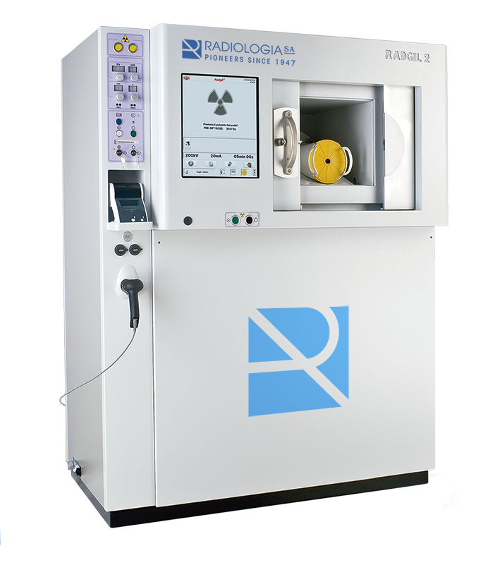
The RADGIL2 X-ray irradiator is a stand-alone X-ray cabinet.
stand-alone X-ray cabinet. It is particularly suitable for
irradiation of blood and blood products to prevent transfusion-associated
prevent transfusion associated graft versus host disease (TA-GVHD), following
host disease (TA-GVHD), following the guidelines of the
(EDQM) to the preparation, use and quality assurance of BLOOD COMPONENT
quality assurance of BLOOD COMPONENT.
View product
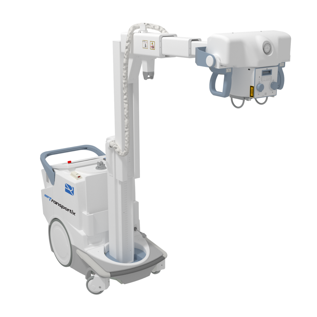
Motorized Digital Battery Mobile, with telescopic column.
View product
 Español
  English