


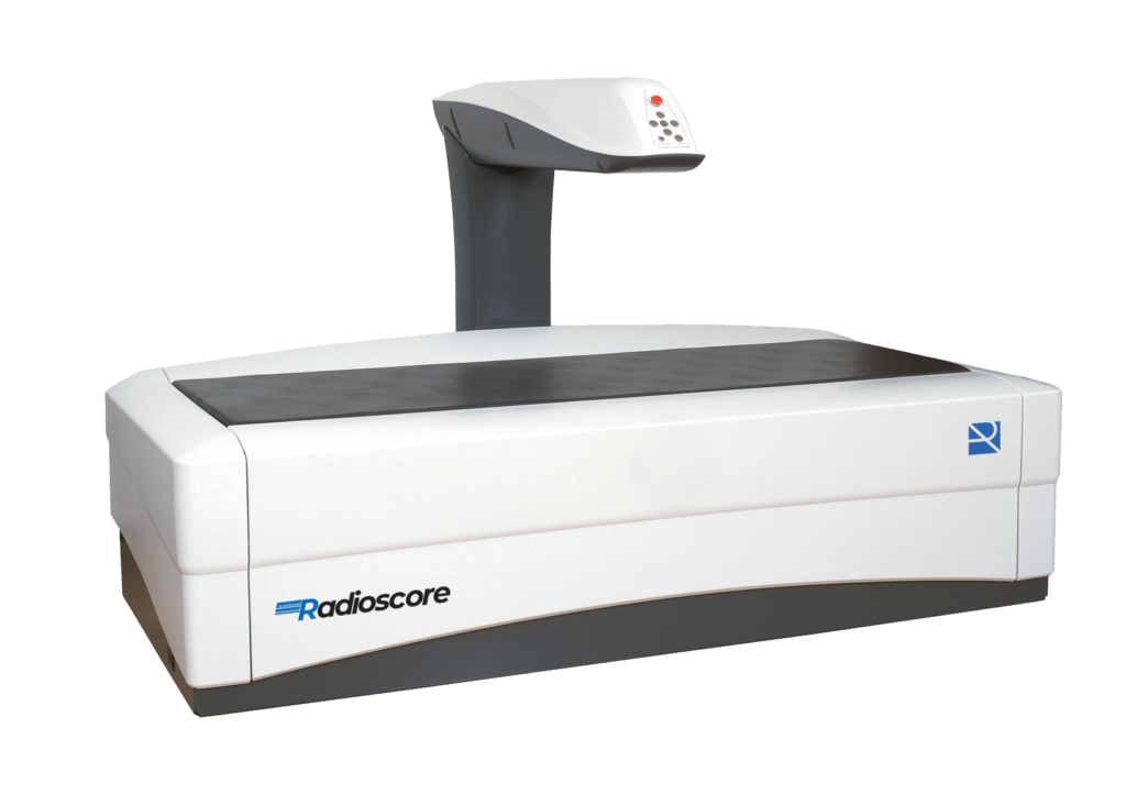
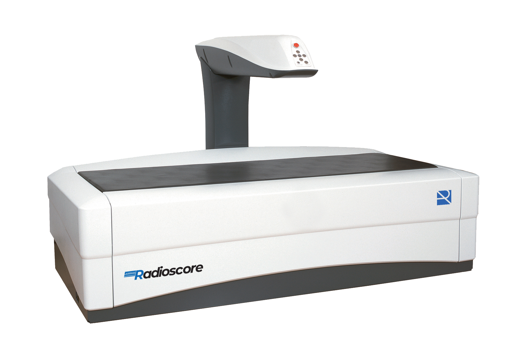
Pencil Beam analog technology DXA bone densitometer, designed for accurate total and regional bone density diagnostics. It allows reliable studies of the main anatomical sites for the evaluation of bone structure and fracture risk, offering an efficient, robust and cost-effective solution for routine clinical practice.
Features
- Complete DXA (Dual-Energy) Densitometry System, designed for routine bone density studies with high diagnostic reliability and optimal patient comfort. It incorporates Pencil Beam Digital Fast technology, an evolution of the traditional Pencil Beam, which allows fast scans with excellent image quality and high measurement accuracy.
- The system offers an optimized workflow thanks to its intuitive software, facilitating patient analysis, diagnosis and follow-up. Its compact design allows installation in small spaces, making it an efficient, robust and cost-effective solution for daily clinical practice.
New outreach to centers and patients
- Diagnosis of osteoporosis by high-precision DXA studies.
- Determination of fracture risk, with analysis of the main anatomical sites (spine and femur).
- Pediatric studies, oriented to growth monitoring and bone age assessment.
- Orthopedic studies, with measurement of bone density in regions of interest and in areas adjacent to prosthetic material.
- Routine clinical evaluation, ideal for centers that require a reliable, efficient and cost-effective solution in bone densitometry.
Full body studies:
- Evaluation of bone density in key anatomical sites: lumbar spine, femur (single or dual) and forearm.
- Precise analysis of regions of interest (ROI), with automatic selection and possibility of manual adjustment by the operator.
- Pencil Beam Digital Fast technology, optimized for fast scans with excellent image quality and high reproducibility.
- Intuitive management and analysis software, which allows control of the examination, evaluation of results and generation of clinical reports.
- Optimized workflow for routine DXA studies with maximum operational efficiency.
- Compact system design, suitable for installations with space limitations.
- Dedicated accessories: anatomical positioners and calibration phantoms.
- DICOM compatibility, for integration with HIS / RIS / PACS systems.
Acquisition
- Low Dose Fluoroscopy.
- High Quality Fluoroscopy (high quality).
- Digital Radiography (instant image).
- Fluoroscopy in Road Mapping mode (optional).
- Fluoroscopy in DSA mode: Remasking, Subtraction, Landmarking, vascular tracking and stenoisis (optional).
Optional
- Upgrade 20kW
- Laser locator.
- Integrated dosimeter.
- DSA vascular packete. In
- Automatic login.
- DICOM functions.
- Wi-Fi pedal.
- RFID card reader.
- Wireless trolley.
- Detector 30×30.
- Rotating anode.
Clinical Use
- Total and regional bone mineral density (BMD) in the main anatomical sites.
- Automatic calculation of T-score and Z-score for the diagnostic evaluation of osteoporosis and osteopenia.
- Analysis of lumbar spine (AP), single and dual femur and forearm, according to clinical indication.
- Fracture risk assessment to support therapeutic decision making.
- Pediatric studies, aimed at monitoring bone growth and bone age.
- Orthopedic applications, with bone density analysis in regions of interest (ROI) and around implants.
- Follow-up of the patient's evolution, using longitudinal analysis and comparison tools.
Technical Specifications
- AP COLUMN
- L1 - L4 (scoliotic analysis). Bone mineral density (BMD), Bone mineral content (BMC), T-score, Z-score, Area.
- ANTEBRAZO
- Ultradistal, Medium-Radius & Distal 1/3. BMD, BMC, T-score, Z-score, Area.
- FEMUR AND DUAL FEMUR
- Total hip, femoral neck, trochanter, intertrochanter, Ward's area. BMD, BMC, T-score, Z-score, Area, Combined left/right double femur & Combined right/left femur exam.
- ONLINE
- Combined examination of lumbar spine and left/right femur. BMD, BMC, T-score, Z-score, Area.
- LATERAL SPINE
- Measurement of bone density of lumbar vertebrae at a lateral angle without spine. BMD, BMC, T-score, Z-score, Area.
- PEDIATRICS
- AP Spine, femur. BMD, MIS, area, skeletal age comparison, Z score.
- ORTHOPEDICS
- Implants in hand, forearm, elbow, shoulder, spine, femur, AP knee, lateral knee, feet. Automatic ROI detection. Bone mineral density (BMD), bone mineral content (BMC), Area.
Documentation
Related Products
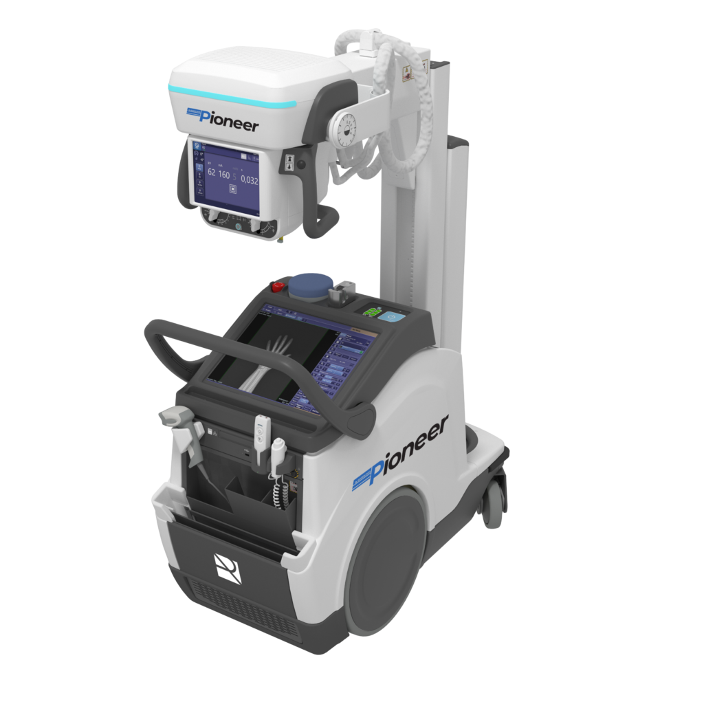
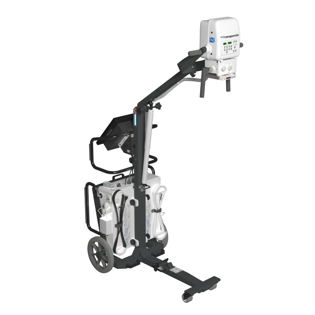
Digital mobile with energy recovery technology, in a lightweight, motorized and compact design that allows you to bring the diagnosis with the patient and more complicated environments such as ICU, neonatal centers, etc..
View product
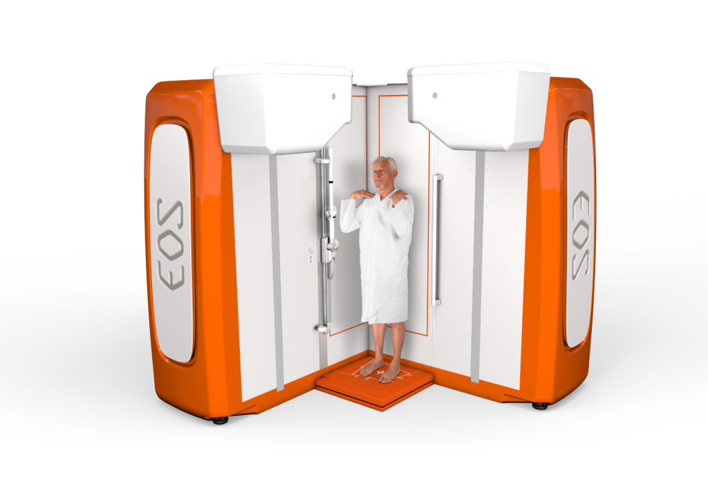
EOSedge proporciona, en un entorno abierto y confortable para el paciente, una evaluación global del sistema esquelético para facilitar la toma de decisiones terapéuticas cruciales.
Los modelos 3D y los parámetros clínicos 3D asociados ayudan a los médicos a evaluar las patologías musculoesqueléticas e identificar opciones de tratamiento para mejorar los resultados clínicos.
View product
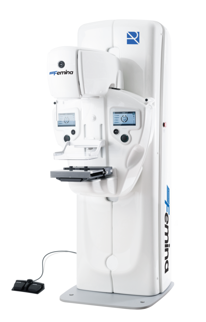
Analog mammography system designed for screening and diagnostic processes, with high frequency generator, Potter-Bucky system and a wide range of accessories for magnification, two-dimensional biopsies and precise positioning, guaranteeing high quality images and reliable diagnosis.
View product
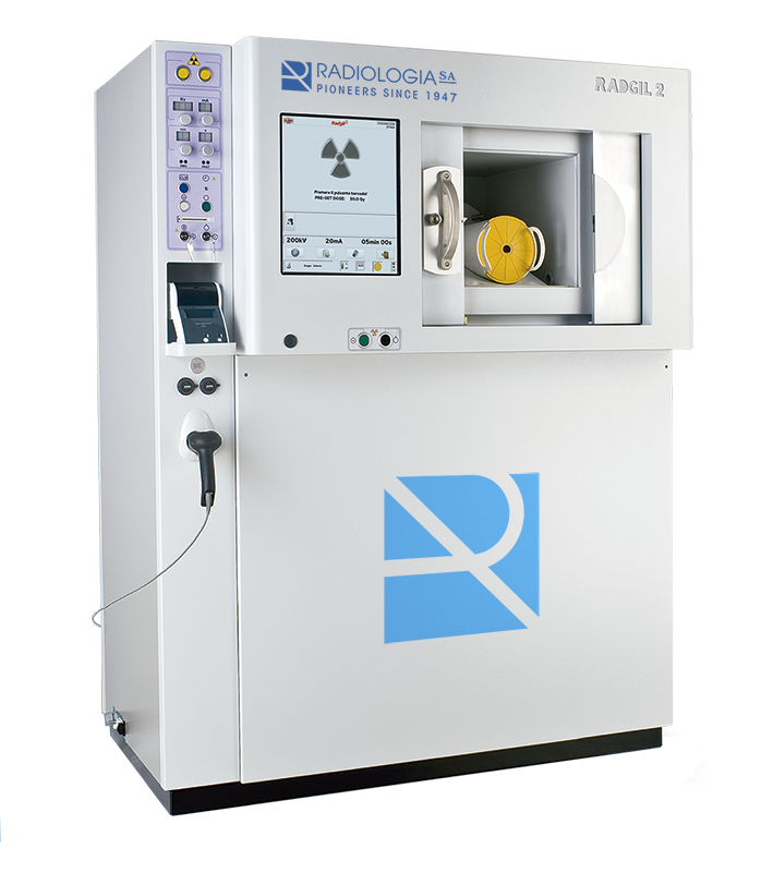
The RADGIL2 X-ray irradiator is a stand-alone X-ray cabinet.
stand-alone X-ray cabinet. It is particularly suitable for
irradiation of blood and blood products to prevent transfusion-associated
prevent transfusion associated graft versus host disease (TA-GVHD), following
host disease (TA-GVHD), following the guidelines of the
(EDQM) to the preparation, use and quality assurance of BLOOD COMPONENT
quality assurance of BLOOD COMPONENT.
View product
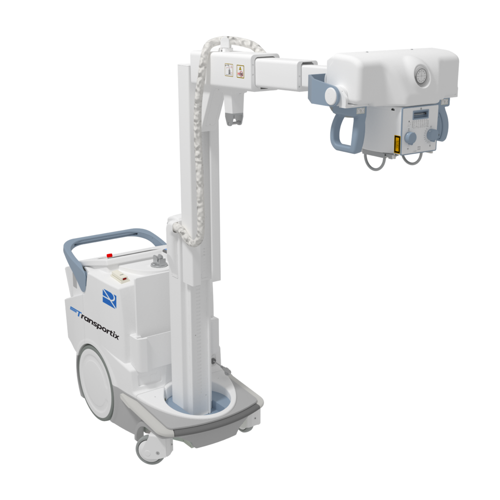
Motorized Digital Battery Mobile, with telescopic column.
View product
 Español
  English